Blog

Hip Abduction / ER Assessment

in Blog

Hip Abduction / ER Assessment

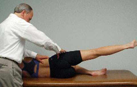
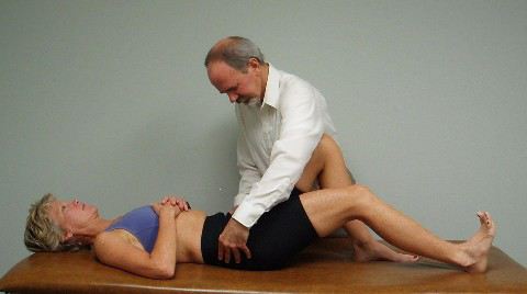
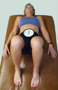
Hip abduction in supine lying with the hips and knees flexed has been commonly given to patients to help strengthen their abdominals while minimizing loading of the low back. Patients have typically been instructed to either flatten their low back while doing this exercise or to find and maintain a neutral lumbar spine during this movement. Most patients are simply instructed to flatten and then extend the low back and find the midpoint in between to obtain what they think is a neutral lumbar spine. Historically, therapists have not paid attention to the stability of the pelvis during this exercise. Mark found that when he placed his thumbs under both ASISs before the start of this exercise that the ASISs were most often asymmetrical to begin with, noting a predominance of the R ASIS appearing inferior when compared to the L side. When this asymmetry is present before initiating the HIP ABDUCTION/ER FUNCTIONAL MOVEMENT TEST, the patient is instructed to level their ASISs by bringing the R side superiorly and then hold the ASISs level as the hips are abducted. He frequently found that patients were unable to maintain the leveling of the ASISs, with the R ASIS usually dropping inferiorly before the L side. He surmised that this is indicative of an imbalance in muscle tone between the hip adductors and the abdominals on the R side signifying a loss of pelvic stability.

Mark also noted during his initial biomechanical evaluation that these patients often presented with an inferior pube dysfunction on the R side. He surmised that if the patient was given the Hip Abduction/ER exercise without consciously maintaining leveling of the ASISs the patient was reinforcing this pelvic dysfunction. Therefore, it is essential that the therapist monitors the ASISs during the performance of this movement test and teaches the patient to limit the degree of hip abduction during retraining based upon their ability to consciously maintain leveling of the ASISs throughout the exercise.

In Rehab Links Module #5 you will learn how to specifically examine your subject using the supine Hip Abduction/ER movement test. In Rehab Links Module #6 the causes and manual treatment to address the premature movement of the pelvis during this movement test are covered in detail. This is followed by the appropriate retraining exercises in Rehab Links Module #7 that are given to patients so that they can eventually perform the Hip Abduction/ER exercise without moving through the pelvis.

Lisa Chase and Mark Bookhout launched the Rehab Links Systems online platform – a unique exercise software for medical, health and fitness professionals – to help users link manual therapy treatments with a client-specific individualized home exercise program that results in improved clinical outcomes. 

Click here to view the online module series and learn more. If you have already taken an exercise course with Bookhout Seminars or Back 2 Normal Institute, Rehab Links also enables you to deepen your knowledge through membership and mentoring opportunities.

Hip Abduction Assessment

in Blog

Hip Abduction Assessment

Hip abduction in side lying with the hips and knees straight is a movement test designed to assess a subject’s ability to activate their gluteus medius muscle in conjunction with the tensor fascia latae and quadratus lumborum. It is believed that the primary role of the gluteus medius is to stabilize the head of the femur in the acetabulum during gait and during active hip abduction, acting biomechanically in a similar fashion as the rotator cuff of the shoulder when elevating the arm. Inhibition/weakness of the gluteus medius is commonly observed and has been reported in patients with a Trendelenburg gait and DJD of the hip. It is therefore essential to assess for the recruitment and activation of the gluteus medius in patients presenting with hip and low back pain symptoms. Dr. Vladimir Janda originally described facilitation of the TFL and/or quadratus lumborum in substitution for gluteus medius inhibition/weakness, resulting in a faulty hip abduction muscle firing pattern. Faulty muscle firing patterns during the HIP ABDUCTION TEST can result in hip hiking by the quadratus lumborum and/or flexion and IR of the leg by an overactive/facilitated TFL.

Mark noted that patients with gluteus medius weakness often presented with a superior and anteriorly displaced humeral head with restricted inferior and posterior glide. He also found a number of other biomechanical dysfunctions contributing to the inhibition of the gluteus medius, including hypertonicity of the hip adductors, lower lumbar segmental dysfunctions involving L4-5 or L5-S1, and/or sacroiliac joint dysfunction on that side. 

In Rehab Links Module #5 you will learn how to specifically examine your subject using the side lying HIP ABDUCTION MOVEMENT TEST. In Rehab Links Module #6 the causes and manual treatment to address the dysfunctional biomechanical factors that result in gluteus medius inhibition are covered in detail. This is followed by the appropriate retraining exercisesin Rehab Links Module #7 that can be given to patients so that they can eventually perform Hip Abduction with the appropriate activation of the gluteus medius and avoid excessive recruitment of the TFL and quadratus lumborum.

Lisa Chase and Mark Bookhout launched the Rehab Links Systems online platform – a unique exercise software for medical, health and fitness professionals – to help users link manual therapy treatments with a client-specific individualized home exercise program that results in improved clinical outcomes. 

Click here to view the online module series and learn more. If you have already taken an exercise course with Bookhout Seminars or Back 2 Normal Institute, Rehab Links also enables you to deepen your knowledge through membership and mentoring opportunities.

Heel Slide How-To

in Blog

Heel Slide How-To

Supine heel slides are commonly given to patients to help strengthen the abdominals and protect the low back. Patients have typically been instructed to either flatten their low back while doing this exercise or to find and maintain a neutral lumbar spine during the heel slide. Most patients are simply instructed to flatten then extend the low back and find the midpoint in between to obtain a neutral lumbar spine. The argument for maintaining a neutral lumbar spine versus flattening the low back is that by simultaneously engaging the posterior musculature to hold a neutral spine it will promote core stability. Historically, therapists have not paid attention to the stability of the innominates during the heel slide exercise. Mark found that when he placed his thumbs under both ASISs at the start of this exercise, the ASISs were most often asymmetrical to begin with, noting a predominance for the R ASIS appearing inferior to the L. He also noted that when monitoring the ASISs during the heel slide movement test, patients often did not keep the ASISs level, with the R ASIS often dropping inferiorly during R heel slide. This is indicative of anterior innominate rotation occurring in substitution for hip extension. 

Mark also noted during his initial biomechanical evaluation that these patients presented with an anteriorly rotated R innominate. He surmised that if the patient was given the heel slide exercise without consciously maintaining leveling of the ASISs the patient was reinforcing this pelvic dysfunction. Therefore it is essential that the therapist monitors the ASISs during the performance of this movement test and teaches the patient to limit the degree of heel slide based upon their ability to maintain leveling of the ASISs. 

In Rehab Links Module #5 you will learn how to specifically examine your subject using the supine heel slide test. In Rehab Links Module #6 the causes and manual treatment to address the premature movement of the innominate during this movement test are covered in detail. This is followed by the appropriate retraining exercises in Rehab Links Module #7 that are given to patients so that they can eventually perform the heel slide exercise without substituting with innominate rotation.  

Lisa Chase and Mark Bookhout launched the Rehab Links Systems online platform – a unique exercise software for medical, health and fitness professionals – to help users link manual therapy treatments with a client-specific individualized home exercise program that results in improved clinical outcomes. 

Click here to view the online module series and learn more. If you have already taken an exercise course with Bookhout Seminars or Back 2 Normal Institute, Rehab Links also enables you to deepen your knowledge through membership and mentoring opportunities.

Perfect the Pelvic Clock

in Blog

Perfect the Pelvic Clock

Mark originally learned the Pelvic Clock from a Feldenkrais practitioner as an Awareness through Movement lesson over 30 years ago and started using it with his low back pain patients to try to improve their sensory awareness and spinal mobility. He discovered that by monitoring the ASISs with his thumbs during pelvic clocks, most patients moved asymmetrically when rolling their pelvis towards 6 or 12. This asymmetrical motion of the ASISs correlated with his biomechanical findings of an associated innominate rotation dysfunction. He then noted that by simply asking the patient to consciously maintain leveling of the ASISs when moving to 12 and 6 that the patient could self treat their innominate dysfunction without the need for manual correction. Also, he found that patients often reported reproduction of their low back pain when attempting to move the pelvis back and forth between 3 and 9 o’clock, and initially that seemed to correlate with a sacroiliac joint dysfunction.  

In addition, by comparing his Osteopathic based biomechanical exam of the lumbar spine to a patient’s restricted mobility with full circle pelvic clocks, he could diagnose lumbar spine non-neutral dysfunctions with remarkable accuracy. For example, a restriction in mobility towards 1-2 o’clock is indicative of an ERS L dysfunction whereas a restriction towards 7-8 o’clock is indicative of an FRS R dysfunction somewhere in the lumbar spine. The ability to diagnose non-neutral lumbar spine dysfunctions is important, as these dysfunctions can result in muscle hyper or hypotonicity and be major players in perpetuating muscle imbalances in the lower quarter.

Rehab Links System Modules 5, 6, and 7 teach how to specifically examine a subject using the pelvic clock, how to interpret the findings, and provide subject specific exercises that are designed to restore a symmetrical full circle active pelvic clock without limitation or provocation of pain. 

Click here to view the online module series and learn more about how to use the Pelvic Clock to make a diagnosis of the lumbar spine and pelvis, and explore the proven treatment and exercise strategies. If you have already taken an exercise course with Bookhout Seminars or Back 2 Normal Institute, Rehab Links also enables you to deepen your knowledge through membership and mentoring opportunities. 

Lisa Chase and Mark Bookhout launched the Rehab Links Systems online platform – a unique exercise software for medical, health and fitness professionals – to help users link manual therapy treatments with a client-specific individualized home exercise program that results in improved clinical outcomes.