Manual Stretching

Dysfunctional Supine Heel Slide Test

in 4 Manual Therapy Strategies, All Manual Therapy, Manual Stretching

Dysfunctional Supine Heel Slide Test

Anatomy and Biomechanics:

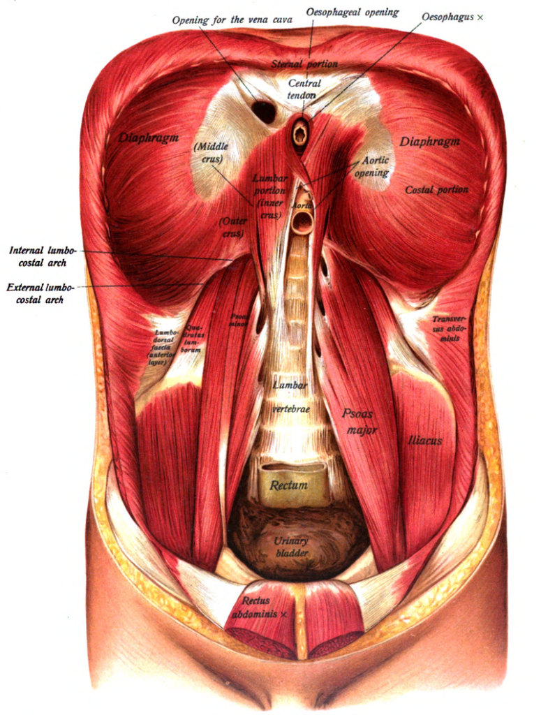
The ability to maintain a stable pelvis (level ASISs) while performing this functional movement test requires normal hip extension ROM and flexibility of the hip flexors with appropriate tone in the abdominal muscles. The abdominals must be strong enough to counterbalance the eccentric recruitment of the iliopsoas as the hip and leg are extended. An imbalance is seen when a hypertonic iliopsoas and weak abdominals results in an ASIS dropping inferiorly during the heel slide test.

After addressing any tightness in the anterior hip capsule, lengthening the hip flexors and mobilizing the femoral nerve as presented in module #3, then proceed to retraining the abdominals.

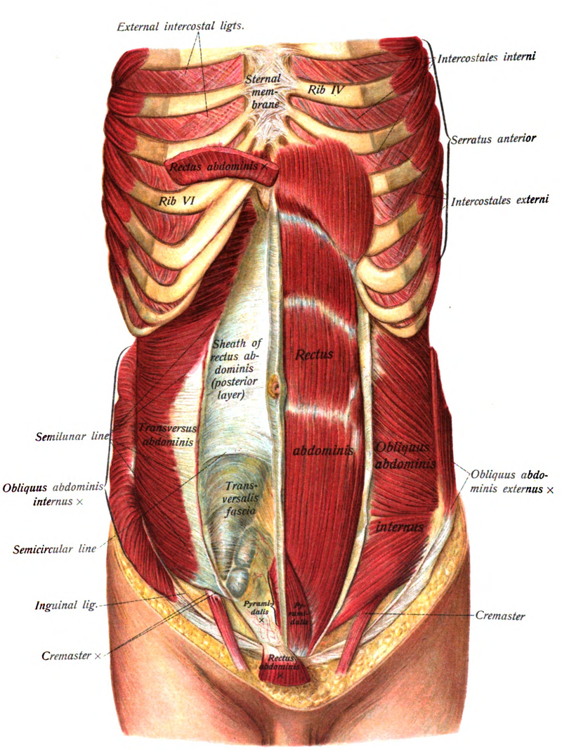
The Abdominals and Multifidi:

Global mobilizer in the sagittal plane – rectus abdominis
Global mobilizer in the transverse plane– internal and external obliques
Segmental stabilizer in the transverse plane – multifidi
Segmental stabilizer in all three planes – transversus abdominis

Rectus Abdominis:

Global mobilizer – brings the pelvis towards the trunk or the trunk towards the pelvis.
This muscle does not contribute to core stability.
Innervation – 7th thru 12th intercostal nerves

Transversus Abdominis:

  • First trunk muscle to normally fire before you move your leg or arm in any direction, but in patients with a history of low back pain the transversus muscle fires late during movement of the lower limb (Hodges and Richardson, 1998) or upper limb (Hodges and Richardson, 1999).
  • The transversus appears to fire as part of a feedforward movement synergy pattern, but contrary to initial studies activation of this muscle is dependent on the direction of movement and often is not bilaterally symmetrical, (Allison, Morris and Lay, 2008)
  • Innervation – 7th thru 12th intercostal nerves, iliohypogastric and ilioinguinal nerves

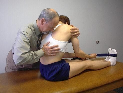
Costovertebral Joint Mobilization with Long Sitting Slump Stretch

in 2 Movement Tests Lower Quarter, 4 Manual Therapy Strategies, All Lower Quarter, All Manual Therapy, Manual Stretching, Mobilization

Costovertebral Joint Mobilization with Long Sitting Slump Stretch

Treatment for Regional Pain Syndrome involving the Right Leg (Adapted from Cleland and McRae, 2002).:

  • Patient is long sitting with a belt wrapped around the balls of the feet.
  • The patient attempts to straighten the knees as much as possible and pull the toes up using the belt.
  • The patient flexes forward as far as possible and drops the head down.
  • The therapist side bends and rotates the patient to the left and holds the patient in this position.
  • The therapist then applies a PA grade III or IV mobilization to ribs 8 through 12 to mobilize the costovertebral joints from T8-12 on the right side.
  • Patient reports on any relief or change in symptoms in the R leg.

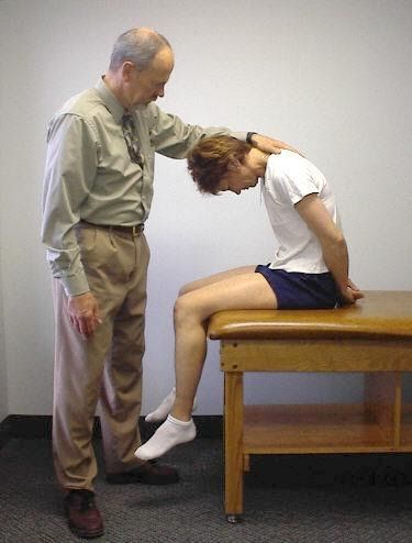
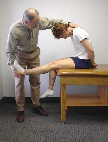
Slump Sitting Mobilization

in 2 Movement Tests Lower Quarter, 4 Manual Therapy Strategies, All Lower Quarter, All Manual Therapy, Manual Stretching, Mobilization, Nerve Glides

Slump Sitting Mobilization

  • Patient sitting at the edge of the table with the neck and trunk flexed, arms behind the back.
  • Therapist stabilizes the head and neck in flexion and passively straightens the leg to the barrier.
  • The therapist then immediately releases the tension by bending the knee.
  • Therapist repeatedly straightens and bends the knee 8-10 times.
  • Patient is then instructed to sit up tall and rock the pelvis forward (6:00) and backward (12:00) to release any tension in the low back.
  • Repeat this sequence with the other leg and then with both legs.
  • Further mobilization can be done by holding dorsiflexion of the ankle while straightening of the knee.

Piriformis Stretch Above 90 Degrees of Hip Flexion

in 2 Movement Tests Lower Quarter, 4 Manual Therapy Strategies, Active Trunk Rotation, All Lower Quarter, All Manual Therapy, Manual Stretching, Muscle Energy, Rolling

Piriformis Stretch Above 90 Degrees of Hip Flexion

  • Therapist externally rotates the leg, then adducts the leg towards the opposite shoulder
  • Patient is instructed to push out with both the knee and ankle; if external rotation is more limited, push out with the ankle only
  • Upon relaxation the therapist adds further adduction and/or ER to the new barrier
  • Repeat 3 to 4 times

Piriformis Stretch Below 90 Degrees of Hip Flexion

in 2 Movement Tests Lower Quarter, 4 Manual Therapy Strategies, Active Trunk Rotation, All Lower Quarter, All Manual Therapy, Manual Stretching, Muscle Energy, Rolling

Piriformis Stretch Below 90 Degrees of Hip Flexion

  • Therapist stands on the opposite side of the table and places the left hand on the L ASIS to stabilize
  • Patient’s left foot is placed outside of the right knee
  • Therapist adducts the leg to the barrier
  • Patient attempts to abduct the leg for 5 -7 seconds then relaxes
  • Repeat 3 to 4 times

Hip Diagonals

in 2 Movement Tests Lower Quarter, 4 Manual Therapy Strategies, 5 Home Exercise Strategy, All Home Exercises, All Lower Quarter, All Manual Therapy, Manual Stretching, Mobilization, Muscle Energy, Self-mobilization

Hip Diagonals

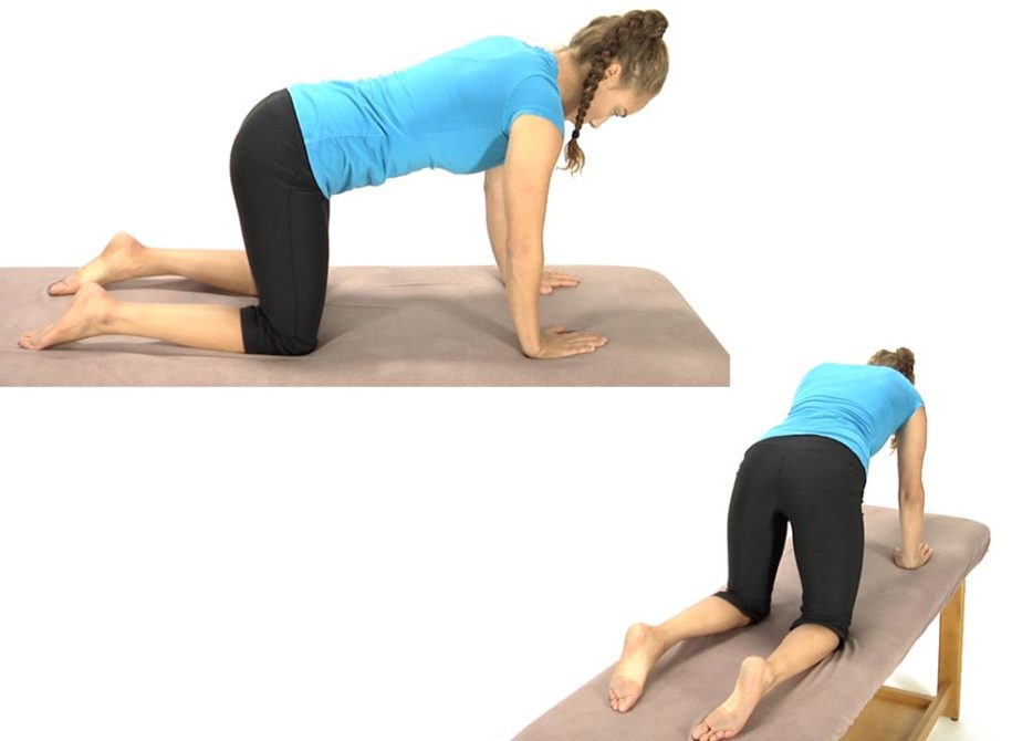
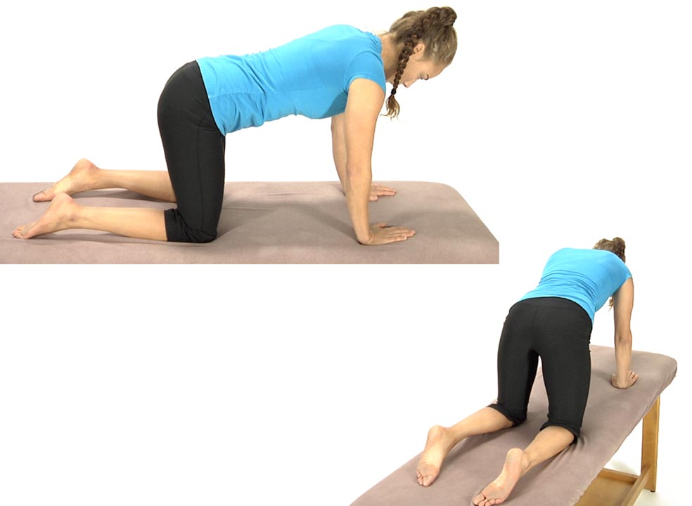
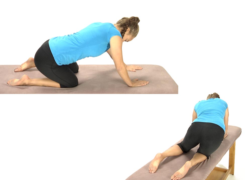
Hip Diagonals

To treat an ERS Right and/or high tone in the Right Erector Spinae:

  • The patient starts this exercise in the hands and knees position and is instructed to sit back diagonally as though attempting to sit onto their right hip.
  • Instruct the patient to sit back as far as they can without holding onto the table with their hands.
  • Have them hold the position for 5 seconds then come back up to the original hands and knees position without pulling up with their arms.
  • Have them repeat 8 to 10 times.

Supine Heel Slide and/or Prone Hip Extension

in 2 Movement Tests Lower Quarter, 4 Manual Therapy Strategies, All Lower Quarter, All Manual Therapy, Manual Stretching

Supine Heel Slide and/or Prone Hip Extension

Hip Flexors

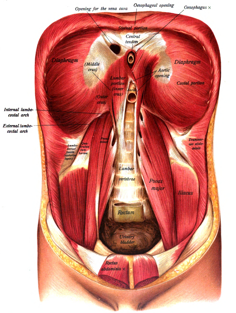
Iliopsoas:

Psoas attaches to the transverse processes and vertebral bodies from T12 to L5
Iliacus attaches to the iliac fossa and sometimes anterior sacral base
Powerful hip flexor and slight ER of the hip, side bends the LS to the same side and rotates away, when hypertonic these muscles limit hip extension
Innervation: Femoral nerve – L2, 3 and 4

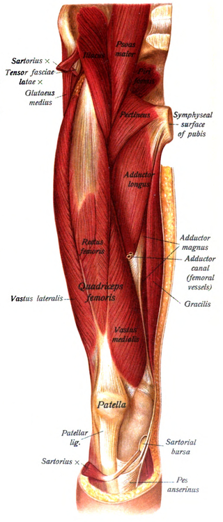
Tensor Fascia Latae:

Attached at the anterior iliac crest and ASIS
Inserts into the IT band
Flexes, abducts and internally rotates the hip, externally rotates the knee
Innervation –Superior gluteal n. L4,5 and S1

Rectus Femoris:

Attached to the AIIS and the anterior hip capsule
Flexes the hip and extends the knee
Pain in the front of the knee cap is often a sign of a tight rectus femoris
Innervation – femoral n. L2, 3, and 4